In addition to being a newly-minted Ph.D., Jenna L. Voellinger is also an exhibiting artist.
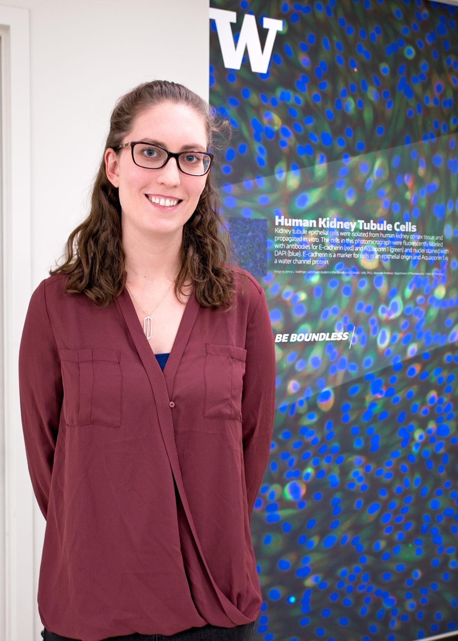
Her image, “Human Kidney Tubule Cells,” created as a graduate student in the laboratory of Edward J. Kelly, Ph.D., Associate Professor, Department of Pharmaceutics, graces one of the classrooms in the T-Wing of the UW Health Sciences Building.
Selected by UW’s design team, Jenna’s image stood out from both a scientific and aesthetic point of view. Her image and those of other researchers are installed in rooms in the T-Wing to inspire students and visitors, while raising awareness of research happening in UW’s Health Sciences.
The image shows kidney tubule epithelial cells that were isolated from human kidney cortex tissue and propagated in vitro. The cells in the photomicrograph were fluorescently labeled with antibodies for E-cadherin (red) and Aquaporin 2 (green) and nuclei stained with DAPI (blue). E-cadherin is a marker for cells of an epithelial origin and Aquaporin 2 is a water channel protein.
To make the image, Jenna used immunocytochemistry to visualize the proteins in the picture. The cells first go through a fixation and permeabilization process that allows the antibodies access to the antigen, or protein, of interest. She then incubates a primary antibody with their cells that is directed against the protein of interest, for example E-cadherin or Aquaporin 2. Next, the cells are incubated with a secondary antibody that attaches to the primary antibody and allows for visualization under a fluorescent microscope. The secondary antibodies have different fluorophores conjugated to them that allow for the different colors seen in the picture, such as green or red.
Jenna successfully defended her dissertation, “Molecular and Cellular Characterization of Human Embryonic Stem Cell Derived Hepatocytes.” Her dissertation research focused on characterizing human embryonic stem cell derived hepatocytes as they relate to primary hepatocytes. This research was been broken into three projects: 1) pharmacogenetic profiling of human embryonic stem cells; 2) characterization of stem cell derived hepatocytes with a focus on CYP-mediated oxidation; and 3) investigating approaches to enhance hepatocyte differentiation focused on the role of miRNAs in the development and maturation of hepatocytes.
“Jenna’s research has been a mixture of biochemistry and cell biology,” said Ed Kelly. “A key component in cell biology research involves microscopy and, as you can see from this image of cultured human kidney cells, the results can be viewed as both art and science.”
Jenna’s art may be seen in T-473.
Interested in applying for an MS or PhD in Pharmaceutics? Click here.